– Report Number: 1-5, – Date: 2024/03/01
Simplified And Modified for Social Media Usage
Referring Veterinarian: Dr. SSS VET Referring Practice: AAA Vet Clinic
Pet owner Mrs./Mr.: E. S., Patient: Jessica, Female Shih Tzu Terrier, 11 years old
Complaint: Lameness and reluctance to move for the past two days.
Requested Radiographic Study: Examination of lumbar spine, pelvis, and right and left hind limbs.
Modality: Radiology
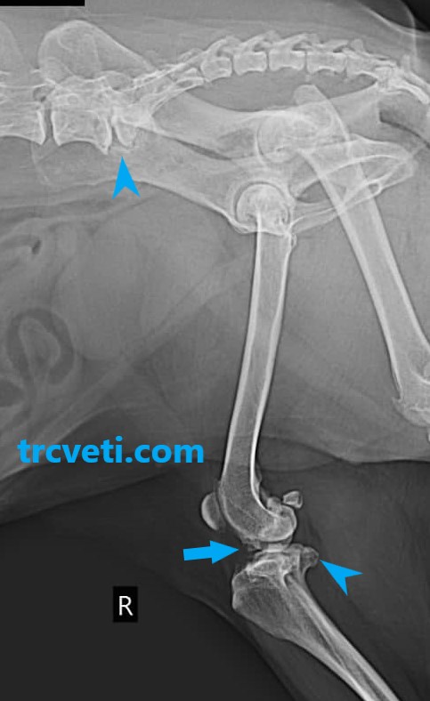
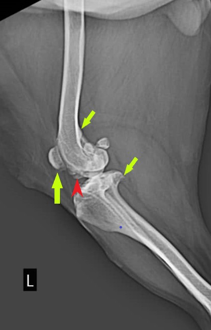
Images: The radiographic study includes abdominal and pelvic views, as well as lateral and VD views of the knee joints. The quality of the radiographs is satisfactory.
Findings:
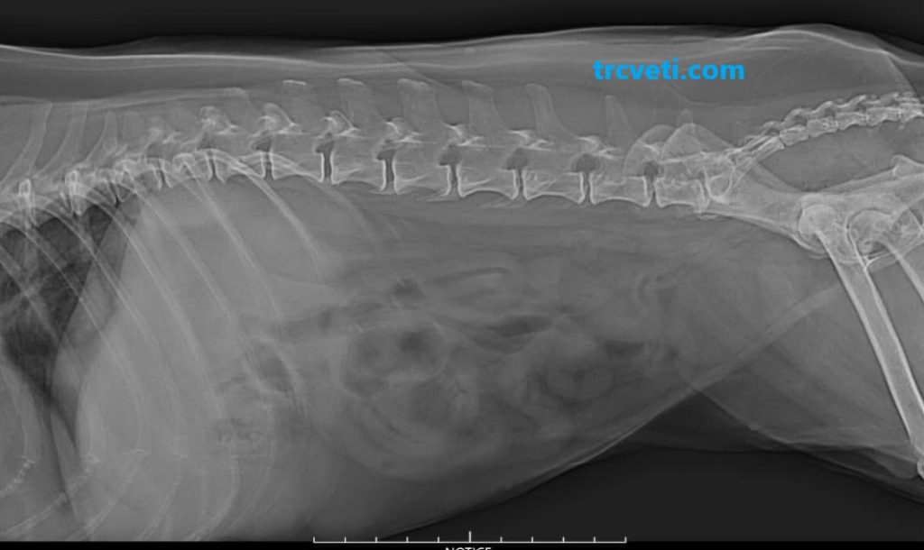
Abdominal Radiographs:
– The kidneys appear smaller than normal and are not well visualized.
– Normal radiographic appearance of the stomach, bowels, urinary bladder, and other abdominal organs.
– No other radiographic abnormalities are detected.
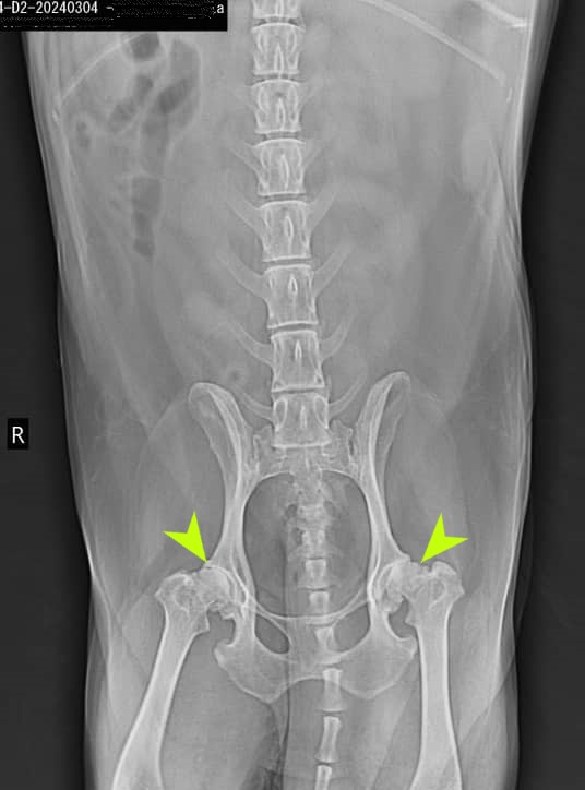
Pelvis and Proximal Hind Limbs:
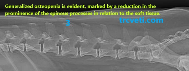
Lumbar Vertebrae:
Note: lumbar vertebrae are included and there is evidence of generalized osteopenia. The spondylosis deformans is seen between L7 and sacrum.
– Radiographic signs of joint disease evident in both hip and knee joints.
– No other abnormalities identified.
Diagnosis and Conclusion:
Abdominal Findings: Normal radiographic appearance with suspicion of renal disease.
Lumbar Vertebrae: Mild renal secondary hyperparathyroidism and spondylosis deformans.
Pelvis and Proximal Hind Limbs: Sever Osteoarthritis of the right and left Hip Knee Joints.
Recommendations:
Performing abdominal ultrasonography is essential and recommended.
Please feel free to contact me for further questions or consultation.
Prepared by a Veterinary Radiologist
ACVRs/EAVDs Diplomates